What is it?
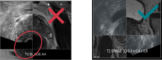
During the MRI/US fusion, on the axial and sagittal views, US and MRI prostate contours are not continuous and don't seem to be the same prostate image.
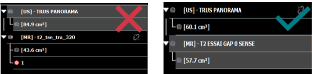
One way to identify incorrect contour is by using the volume measurement, enabled with the option Promap™ VM. The maximum volume difference accepted is 15%:
How to investigate?
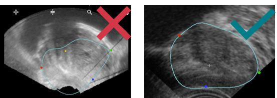
- Check the location of the 3 main points:
-
- Apex in green
- Bladder neck in red
- Median posterior in blue
-
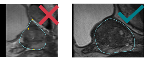
- Make sure the blue contour fits correctly with the prostate shape on MRI & US:
- At the end of your investigations, if none of the steps above solved the issue retrieve the patient data and open a customer request via this form. Attach the data to the form.
How to solve?
In the vast majority of the cases, the issue is due to a misuse and one of the steps above will solve the issue. Otherwise, KOELIS Service team will perform deeper investigations and will come back to you quickly.